
Negative-pressure wound therapy can be a remarkably powerful tool in in the treatment of complex wounds, including diabetic foot ulcers. The keys to success with negative-pressure wound therapy are patient selection, wound preparation, and device application. In this article, we review the mechanism of action and clinical applications of negative-pressure wound therapy laid out by Hasan et al.1
How negative-pressure wound therapy works
Much of what we know about negative-pressure’s mechanism of action comes from the work of Morykwas in animal models.2 Negative-pressure wound therapy is believed to create an optimal wound healing environment by promoting or facilitating several conditions within the wound bed, including:
Local blood flow enhancement: Negative-pressures increase blood flow to wound tissues, presumably by acting through vasomotor mediators. The generally agreed-upon pressure level for this purpose is -125 mmHg, though pressures up to -300 mmHg may confer additional benefits.3
Macrodeformation: Negative-pressure wound therapy facilitates wound contraction through macrodeformation, which can help reduce the size of larger wounds.
Granulation tissue formation and angiogenesis: Negative-pressure wound therapy is superior to traditional dressings in facilitating the formation of granulation tissue. Moreover, microdeformation stimulates vasculogenesis, particularly angiogenesis.
Local fluid reduction: Negative-pressure wound therapy may be particularly helpful in wounds that produce copious amounts of exudate. This intervention reduces fluid from the wound and local edema.4
While some authors have suggested that negative-pressure wound therapy reduces bacterial colonization5, this effect seems to be no more sophisticated than simple barrier protection.6
Choosing the right patient for negative-pressure wound therapy
Negative-pressure wound therapy should be considered in all patients with necrotizing fasciitis, foot abscesses, and infected heel ulcers with deep wounds (i.e., exposed bone, capsule, or tendon).1 However, not all patients with these wounds will be optimal candidates for this intervention.
Severe ischemia is a contraindication to negative-pressure wound therapy. Indeed, high negative pressures may worsen ischemia. Clinically speaking, at least one pulse should be palpable in the affected foot and capillary refill time must not exceed 2 seconds.1
Deep tissue infection (e.g., osteomyelitis, septic arthritis) is a contraindication to negative-pressure wound therapy. The application of a negative pressure device may effectively create an abscess in which the infection can flourish. Deep tissue infections must be treated separately and aggressively. In fact, negative-pressure wound therapy should only be used after the wound has been radically debrided, removing all necrotic, devitalized, or infected tissues.
Using negative-pressure wound therapy: Tips and Tricks
How do I apply the dressing?
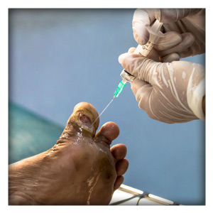
For documentation purposes, a high-resolution photo should be taken of the wound before applying the initial dressing. Keep in mind that the goal of this therapy is to create a negative pressure or vacuum on the wound. Thus, wound preparation and device application are critical. First, the wound edges should be framed with a barrier dressing, and a foam dressing should be placed into the wound defect. An adhesive drape should be secured over the wound allowing a sufficient border on all sides. The drape must make full contact with healthy skin to generate the negative pressure. Adhesive tape should be applied at the border of the dressing to ensure an airtight seal.
A small slit should be made in the center of the filled-in wound, over which the negative pressure tube can be secured and connected to a vacuum pump. If successful, the device should shrink with applied suction with leaks. If a leak is detected, adhesive tape can be used to form a seal.
How often do I change dressings?
The negative-pressure dressing is generally replaced every 3 days. A dressing change is in important time to make assessments and reevaluate therapeutic goals. Compare the initial photo with the current state of the wound to track healing or illness progression. A healing wound should be clean, red, and have sufficient amounts of granulation tissue. If there are overt signs of infection or devitalized tissue, these should be removed via bedside debridement (or surgical debridement, if necessary).
What filler should I use?
The wound may be filled with black foam (polyurethane), white foam (polyvinyl alcohol), or saline-moistened gauze. Black foam is hydrophobic, creates a good phone tissue interface, and promotes rapid, thick granulation.7 Black foam is a good choice for large wounds because it facilitates wound contracture.
White foam is hydrophilic, has higher tensile strength, and is less adherent than black foam. White foam is a good choice for wounds with tunneling or undermined tissue, and for wounds with exposed bone. Saline-moistened gauze has roughly the same characteristics as white foam except with less wound deformation. Clinical results with saline-moistened gauze and white foam are similar, however.8
Silver-impregnated dressings and foams may lessen wound exudate and microbial burden and, consequently, facilitate healing.1 Silver products may offer an advantage to non-silver versions of the same products.
What adhesive tape should I use?
The ideal adhesive tape for negative-pressure wound therapy is hypoallergenic (ideally latex-free), waterproof, and soil-resistant. Moreover, the adhesive tape should fully conform to the body such that it can form a tight seal. The ideal adhesive tape should hold firmly under negative pressure, but it should also be easy to remove during dressing changes every 3 days. Of the adhesive tape options commercially available, Hy-Tape meets or exceeds all of these conditions.
What pressure setting should I use?
The standard initial pressure setting is -125 mmHg. Slightly higher pressures may be acceptable (150 mmHg) for wounds with copious exudate. Lower pressures (50 mmHg) may be reasonable if there is a concern that tissue ischemia may develop.
In most cases, continuous negative pressure mode should be used. While clinical studies have suggested intermittent mode may lead to greater granulation9, patients report greater pain when treated with intermittent or variable negative pressures.1,9
When can I stop negative-pressure wound therapy?
Since the goal of negative-pressure wound therapy is to prepare the wound bed, the device can be removed once this step has occurred. Evidence of wound bed preparation includes a complete base of granulation tissue on all surfaces of the wound, and a wound bed that is free of microorganisms by culture. In unsuccessful cases, negative-pressure wound therapy should be stopped if the wound or distal tissues become ischemic or deep tissue infection develops.
Click Here to Request a Sample of Hy-Tape
References
1. Hasan MY, Teo R, Nather A. Negative-Pressure Wound Therapy for Management of Diabetic Foot Wounds: A Review of the Mechanism of Action, Clinical Applications, and Recent Developments. Diabetic Foot & Ankle. 2015;6:10.3402/dfa.v3406.27618. doi:10.3402/dfa.v6.27618
2. Morykwas MJ, Argenta LC. Nonsurgical Modalities to Enhance Healing and Care of Soft Tissue Wounds. J South Orthop Assoc. 1997;6(4):279-288.
3. Timmers MS, Le Cessie S, Banwell P, Jukema GN. The Effects of Varying Degrees of Pressure Delivered by Negative-Pressure Wound Therapy on Skin Perfusion. Ann Plast Surg. 2005;55(6):665-671.
4. Lu X, Chen S, Li X. The Experimental Study of the Effects of Vacuum-Assisted Closure on Edema and Vessel Permeability of the Wound. Chin J Clin Rehab. 2003;7:1244-1245.
5. Vig S, Dowsett C, Berg L, et al. Evidence-Based Recommendations for the Use of Negative Pressure Wound Therapy in Chronic Wounds: Steps Towards an International Consensus. J Tissue Viability. 2011;20 Suppl 1:S1-18. doi:10.1016/j.jtv.2011.07.002
6. Patmo AS, Krijnen P, Tuinebreijer WE, Breederveld RS. The Effect of Vacuum-Assisted Closure on the Bacterial Load and Type of Bacteria: A Systematic Review. Adv Wound Care (New Rochelle). 2014;3(5):383-389. doi:10.1089/wound.2013.0510
7. Borgquist O, Gustafson L, Ingemansson R, Malmsjo M. Tissue Ingrowth into Foam but Not into Gauze During Negative Pressure Wound Therapy. Wounds. 2009;21(11):302-309.
8. Malmsjo M, Ingemansson R, Martin R, Huddleston E. Negative-Pressure Wound Therapy Using Gauze or Open-Cell Polyurethane Foam: Similar Early Effects on Pressure Transduction and Tissue Contraction in an Experimental Porcine Wound Model. Wound Repair Regen. 2009;17(2):200-205. doi:10.1111/j.1524-475X.2009.00461.x
9. Malmsjo M, Gustafsson L, Lindstedt S, Gesslein B, Ingemansson R. The Effects of Variable, Intermittent, and Continuous Negative Pressure Wound Therapy, Using Foam or Gauze, on Wound Contraction, Granulation Tissue Formation, and Ingrowth into the Wound Filler. Eplasty. 2012;12:e5.